Cardiac function and structure
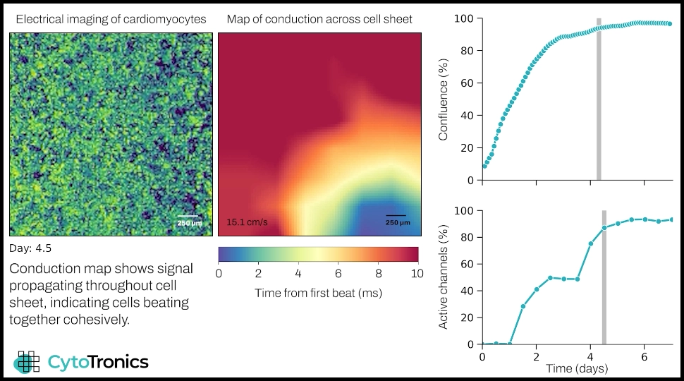
Pixel captures real-time electrophysiology and morphology of cardiomyocytes in one multiplexed assay.
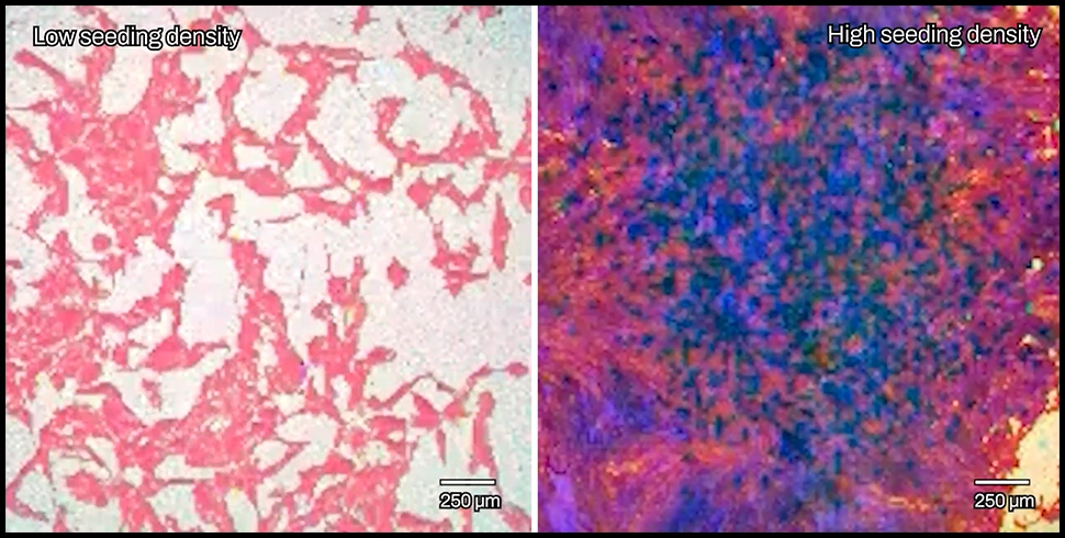
This video shows the maturation of a cardiomyocyte cell sheet, with culture dynamics visible on the left, and propagation of electrical signal throughout the cells visible in the conduction heat map. The conduction map shows how cells initially beat in isolated clumps, while after a few days the signal traverses the culture, indicating the development of a functionally cohesive cell sheet. The plots show culture growth (top) and percentage of beating cells (bottom).