• Pixel
    • Pixel Systems
    • Technology
    • Applications
  • Resources
    • Publications
    • Application Notes
    • Case Studies
    • Videos
    • Blog
  • About
    • Our Story
    • Team
    • News
    • Events
    • Careers
  • Contact
  • Pixel Portal
  • Request a Quote
  • Menu Menu
  • Pixel
    • Pixel Systems
    • Technology
    • Applications
  • Resources
    • Publications
    • Application Notes
    • Case Studies
    • Videos
    • Blog
  • About
    • Our Story
    • Team
    • News
    • Events
    • Careers
  • Contact
  • Pixel Portal
  • Request a Quote

Resources

Information and insights

Resources Type
  • All
  • Application Note
  • Blog
  • Case Study
  • Poster
  • Publication
  • Spec Sheet
  • Technical Note
  • Video
BlogCardiac Function

Cardiac function and structure: Insights for disease models and drug discovery

View >
Technical NoteNeuron Function

Electrophysiological and Morphological Profiling of Human Neurons with the Pixel System

View >
BlogNeuron Function

Neural electrophysiology: Measuring how neurons communicate

View >
BlogEndothelial and Epithelial Cells

Modeling the blood-brain barrier: Tools, challenges, & solutions

View >
Video

Application highlights

View >
Case StudyEndothelial and Epithelial Cells

In vitro modeling of vascular biology using human umbilical vein endothelial cells

View >
Case StudyMoA/Toxicology

Identifying mechanisms of cytotoxicity using electrical imaging

View >
Case StudyMoA/ToxicologyCancer Cell Biology

Comparison of high-dimensional profiling in diverse cell lines

View >
Case StudyEndothelial and Epithelial Cells

In vitro disease modeling in human coronary artery endothelial cells

View >

Contact us

12 Farnsworth St.
Boston, MA 02210
© CytoTronics 2025
Privacy Policy
Vulnerability Disclosure Policy

Stay connected

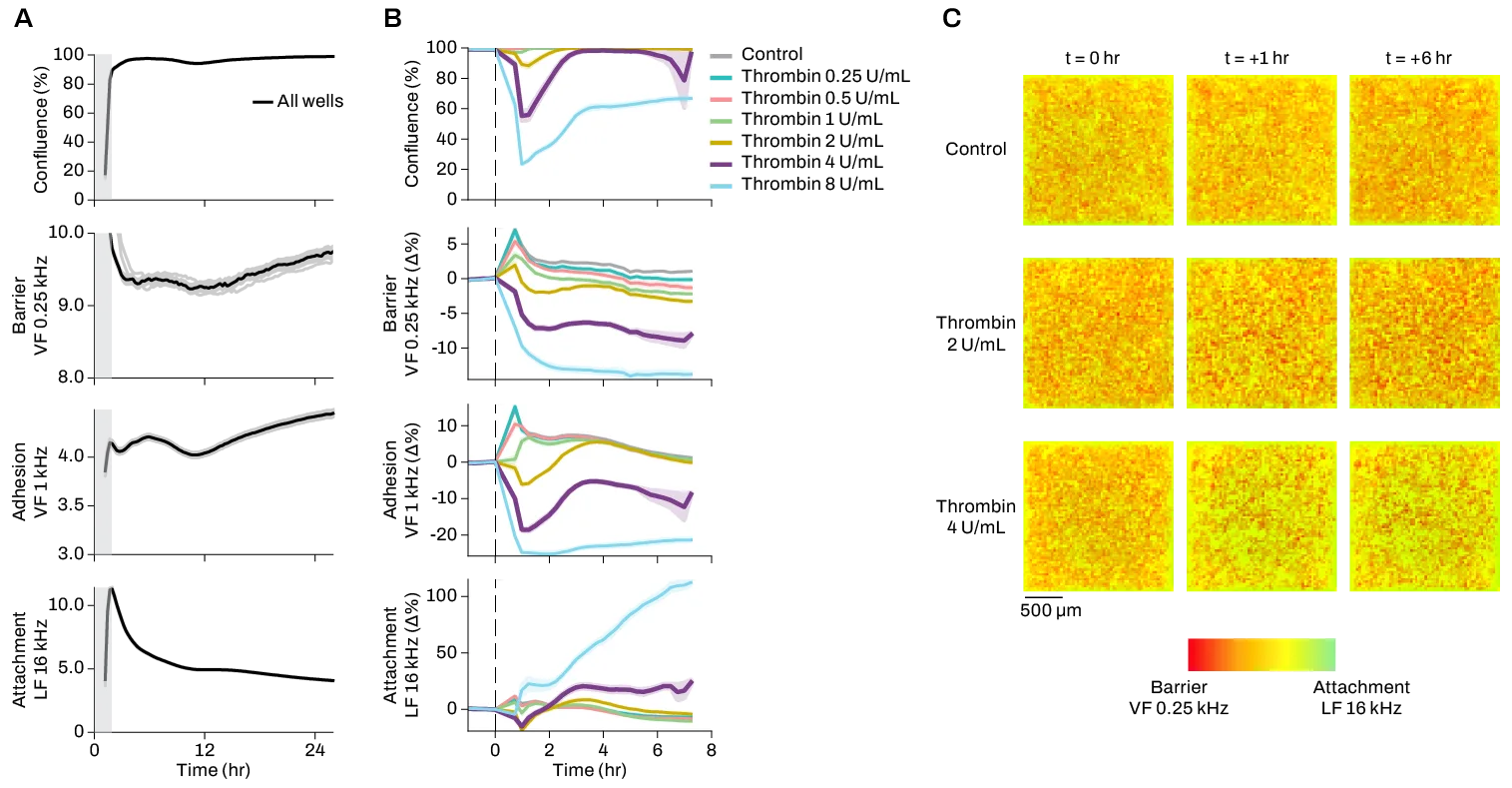
Longitudinal measurements of HUVEC cells upon addition of thrombin (0, 0.25, 0.5, 1, 2, 4, 8 U/mL). (A) Confluence, barrier, cell-cell adhesion, and attachment as measured for 8 hours after thrombin addition as indicated by the dashed line. Shaded regions indicate standard error of 3–6 technical replicates. (B) Electrical images for three conditions (0, 2, and 4 U/mL) prior to thrombin addition (0 hours) and post thrombin addition (+1 and +6 hours) where red represents barrier and green represents attachment.


Request a private Zoom meeting for pre-release product introduction

Entry limit reached.

  • Pixel
    • Pixel Systems
    • Technology
    • Applications
  • Resources
    • Publications
    • Application Notes
    • Case Studies
    • Videos
    • Blog
  • About
    • Our Story
    • Team
    • News
    • Events
    • Careers
  • Contact
  • Pixel Portal
  • Request a Quote
  • Request a Quote